Research
Our research publications are primarily in the domain of cognitive and systems neuroscience. Our research aims to understand the organization of the cerebral cortex through broadly investigating patterns of connectivity in relation to cortical structure. Below are brief summaries of several lines of our research:
→ cortical gradients
→ connectivity visualization
→ manual area parcellation
→ comparative connectomics
→ self-generated thought
→ age-prediction
→ group synchrony
Projects
Gradients in cortical organization
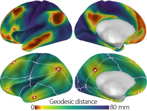
What are the spatial trends underlying the organization of the cerebral cortex, and what are their functional implications? Our primary research agenda focuses on describing these topographic principles of cortical organization. We recently characterized a principal axis of cortical connectivity,1 which relates to cortical geometry and captures a functional hierarchy. We have also mapped the relationship between the principal gradient and measures of cortical microstructure,2 as well as the distance between interconnected areas.3,4
Through ongoing collaborations, we have further probed the functional implications of the principal gradient,5 and investigated its phylogenetic precursors in other primate species, including the marmoset monkey.6
Our future research expands on this axis of connectivity to establish a coordinate system with the aim of differentiating cognitive states, investigating inter-individual variance in brain-behaviour relationships, and mapping individual cortical anatomy.
For an overview, see our recent review article7 and the OHBM keynote presentation.
Gradient maps in HCP 32k cifti, fsaverage, and volumetric spaces can be downloaded here: Gradient templates
-
1 | PDF | SI | Link | Code | | Data | Margulies DS, Ghosh SS, Goulas A, Falkiewicz M, Huntenburg JM, Langs G, Bezgin G, Eickhoff SB, Castellanos FX, Petrides M, Jefferies E, Smallwood J (2016) Situating the default-mode network along a principal gradient of macroscale cortical organization Proc Natl Acad Sci U S A 113(44): 12574–12579
Visualization of cortical connectivity
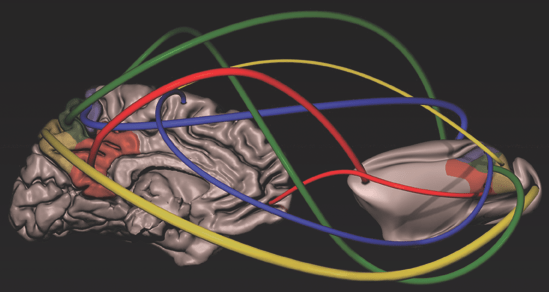
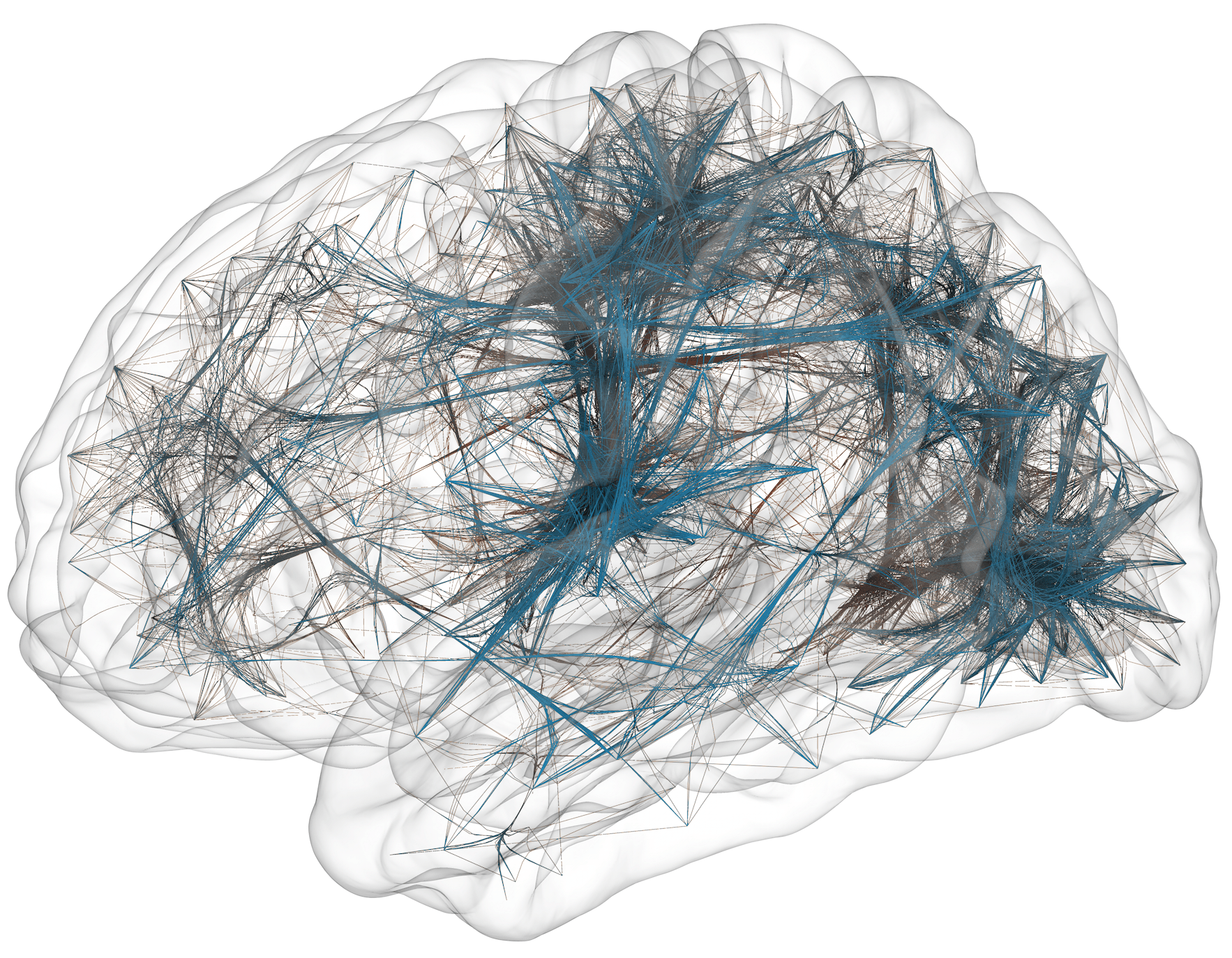 The visualization of large-scale brain connectivity requires specialized tools and consideration of the features of interest.1
Joachim Böttger developed two approaches: edge-bundling2 and connectivity glyphs3, which we applied to the manual parcellation of Broca’s area.
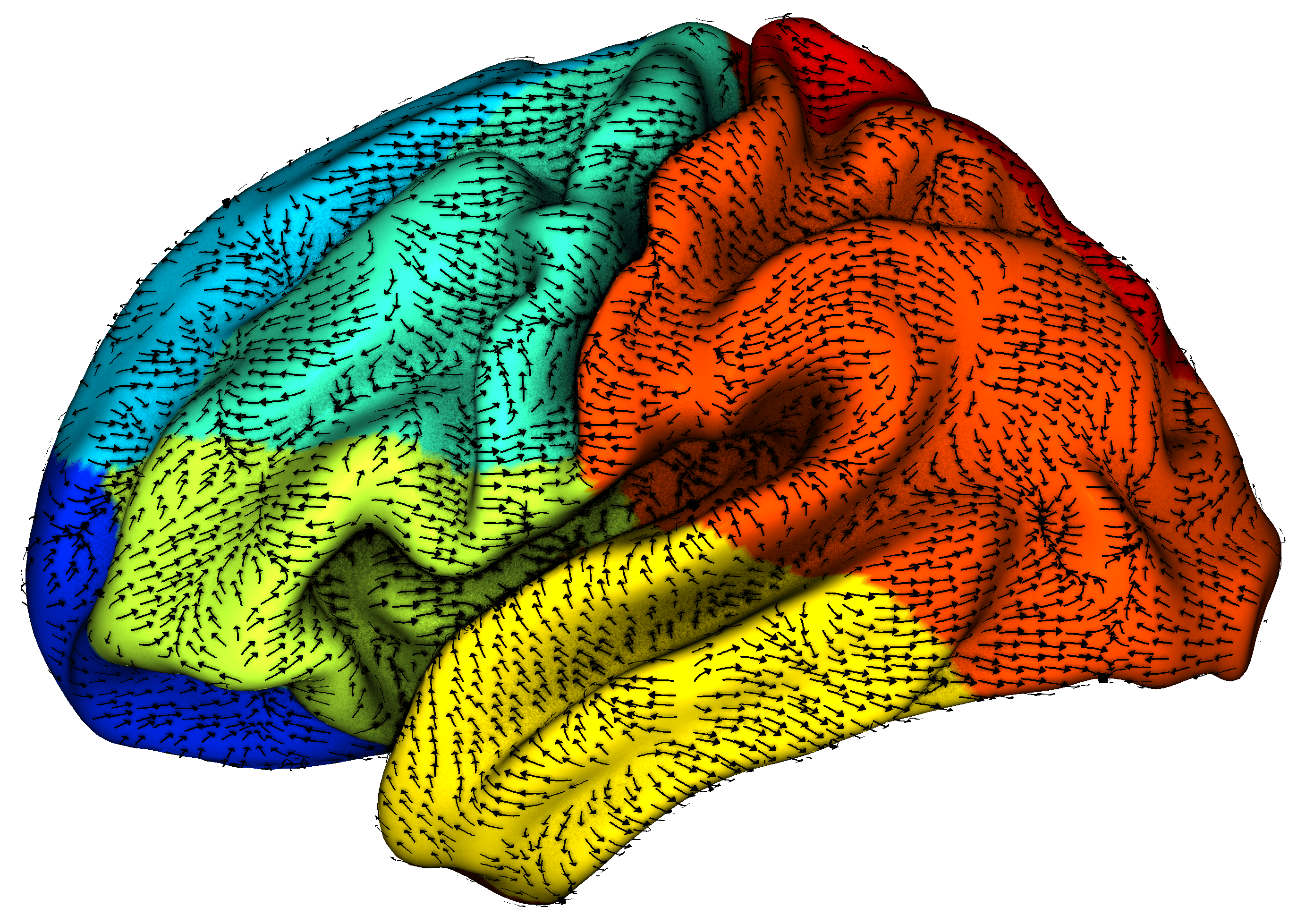
To visualize large-scale spatial trends along the cortical surface, we developed the visualization software Directionality Indicator, which projects arrows onto the cortical surface based on the orientation of streamlines that follow a progression of cortical features.
Manual and Automated Parcellation of Broca’s area
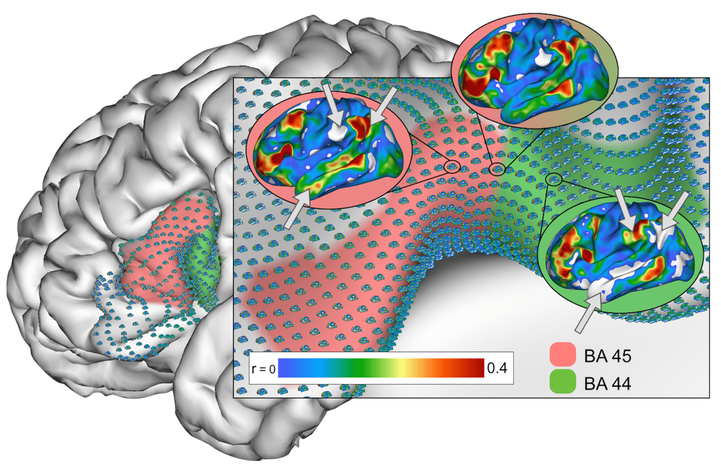 Building on our work describing connectivity-based subdivisions with ventrolateral frontal cortex on the group-1 and individual-level2, we aimed to delineate the extent of areas 44 and 45 at high-resolution. Using novel software tools we developed to visualize high-resolution functional connectivity data, Estrid Jakobsen manually delineated these areas in 101 individual datasets from the Human Connectome Project (HCP)3, and developed an automated pipeline for delineating these areas in further datasets4.
Manually delineated labels for areas 44 and 45 in 101 HCP participates are available here.
Cross-species comparative cortical connectivity
 Comparative studies of connectivity organization between different species enable the translation across literatures. The macaque monkey offers a half-century of gold-standard knowledge of primate cortical connectivity based on tract-tracing studies. We have investigated subdivisons with the posteromedial cortex1, ventral frontal cortex2, as well as parcellation of the macaque monkey lateral frontal cortex3.
In addition, we characterized the principal gradient in marmoset monkeys based on tract-tracing data6.
We have also co-organized the openly available non-human primate MRI database: the Primate Neuroimaging data-exchange (PRIME-DE)5.
-
5 | PDF | Link | | Data | Milham MP, Ai L, Koo B, Xu T, Amiez C, Balezeau F, Baxter MG, Blezer ELA, Brochier T, Chen A, Croxson PL, Damatac CG, Dehaene S, Everling S, Fair DA, Fleysher L, Freiwald W, Froudist-Walsh S, Griffiths TD, Guedj C, Hadj-Bouziane F, Ben Hamed S, Harel N, Hiba B, Jarraya B, Jung B, Kastner S, Klink PC, Kwok SC, Laland KN, Leopold DA, Lindenfors P, Mars RB, Menon RS, Messinger A, Meunier M, Mok K, Morrison JH, Nacef J, Nagy J, Rios MO, Petkov CI, Pinsk M, Poirier C, Procyk E, Rajimehr R, Reader SM, Roelfsema PR, Rudko DA, Rushworth MFS, Russ BE, Sallet J, Schmid MC, Schwiedrzik CM, Seidlitz J, Sein J, Shmuel A, Sullivan EL, Ungerleider L, Thiele A, Todorov OS, Tsao D, Wang Z, Wilson CRE, Yacoub E, Ye FQ, Zarco W, Zhou Y-di, Margulies DS, Schroeder CE (2018) An Open Resource for Non-human Primate Imaging Neuron (100)
Connectomic correlates of self-generated thought
 How does spontaneous brain activity relate to the dynamics of ongoing thought? In collaboration with Jonny Smallwood we have been pursuing this question through a series of studies that combine cognitive and behavioral paradigms, psychological questionnaires, and neuroimaging data.1,2,3,4,5
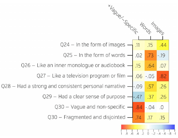
We have also validated a questionnaire that captures the content and form of mind wandering,6 which is openly available here.
-
3 | PDF | Link | Golchert J, Smallwood J, Jefferies E, Seli P, Huntenburg JM, Liem F, Lauckner ME, Oligschlager S, Bernhardt BC, Villringer A, Margulies DS (2017) Individual variation in intentionality in the mind-wandering state is reflected in the integration of the default-mode, fronto-parietal, and limbic networks NeuroImage 146: 226–235
-
4 | PDF | Link | Smallwood J, Gorgolewski KJ, Golchert J, Ruby FJM, Engen H, Baird B, Vinski MT, Schooler JW, Margulies DS (2013) The default modes of reading: modulation of posterior cingulate and medial prefrontal cortex connectivity associated with comprehension and task focus while reading Front Hum Neurosci 7: 734
-
5 | PDF | Link | Smallwood J, Karapanagiotidis T, Ruby F, Medea B, de Caso I, Konishi M, Wang H-T, Hallam G, Margulies DS, Jefferies E (2016) Representing Representation: Integration between the Temporal Lobe and the Posterior Cingulate Influences the Content and Form of Spontaneous Thought PLoS One 11(4): e0152272
Age prediction
 Franz Liem recently developed a multimodal imaging approach to age prediction.1 For a plug-and-play version based on cortical thickness, cortical surface area, and subcortical information, check out Brain-Age Regression Analysis and Computation Utility Software as the BARACUS Bids App
-
1 | PDF | Link | Code | Liem F, Varoquaux G, Kynast J, Beyer F, Kharabian Masouleh S, Huntenburg JM, Lampe L, Rahim M, Abraham A, Craddock RC, Riedel-Heller S, Luck T, Loeffler M, Schroeter ML, Witte AV, Villringer A, Margulies DS (2017) Predicting brain-age from multimodal imaging data captures cognitive impairment NeuroImage 148: 179–188
Group synchrony
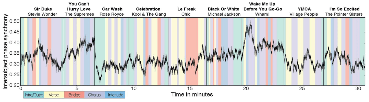 How do individuals synchronize their behavior when in a group?1 Melissa Ellamil investigated this question in a club setting in collaboration with Guerilla Science,2 which was also described in PNAS News. Data from this study are openly available here.